Overview
Percutaneous Nephrolithotomy (PCNL) is a minimally invasive surgical procedure used primarily for the treatment of larger or complex kidney stones.
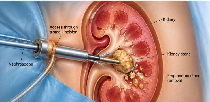
During PCNL, the urologist creates a small access tract through the skin of the back directly into the kidney. A specialized instrument is then introduced through this tract to locate, fragment and remove the stone.
PCNL is particularly useful when a kidney stone is too large or complex to be effectively treated with less invasive stone-treatment approaches.
Common Symptoms
PCNL is a treatment procedure rather than a condition itself. Patients requiring PCNL may have symptoms or findings related to larger or complex kidney stones, such as:
• Severe or persistent flank pain
• Blood in the urine
• Recurrent urinary tract infections
• Difficulty passing urine when obstruction is present
• Urinary obstruction caused by a stone
• Large kidney stones identified on imaging
• Multiple or complex kidney stones
The decision to perform PCNL depends on the stone's size, location, number, composition and the patient's overall condition.
Possible Causes
PCNL may be recommended when kidney stones are large, complex or unlikely to be adequately treated with other stone-removal approaches.
Common reasons for considering PCNL include:
• Large kidney stones
• Staghorn or branching kidney stones
• Multiple kidney stones
• Complex stone burden
• Stones causing significant obstruction
• Stones that are not suitable for or have not responded adequately to other treatments
The appropriate treatment is determined after imaging and specialist assessment.
Diagnosis
Before PCNL, the urologist evaluates the patient's overall health and the characteristics of the kidney stone.
Assessment may include:
• Detailed medical and surgical history
• Physical examination
• Urine examination and urine culture
• Blood tests
• Kidney function assessment
• Ultrasound or X-ray when appropriate
• CT scan to determine stone size, location and complexity
• Assessment of the urinary tract and kidney anatomy
The evaluation helps the urologist plan the safest access route and determine the appropriate treatment approach.
Treatment & Procedures
PCNL is generally performed under anesthesia.
The procedure typically involves:
• Positioning the patient appropriately for the procedure
• Using imaging guidance to access the kidney through a small opening in the back
• Creating a controlled tract into the kidney
• Introducing a nephroscope through the access tract
• Locating the kidney stone
• Fragmenting the stone using an appropriate energy source when required
• Removing the stone fragments through the access tract
• Checking the kidney and urinary drainage at the end of the procedure
Depending on the individual case, a temporary nephrostomy tube or ureteral stent may be placed after the procedure.
The exact technique and postoperative drainage plan depend on the stone burden, anatomy and clinical circumstances.
Recovery & Follow-up
Recovery after PCNL varies depending on the size and complexity of the stone, the procedure performed and the patient's overall health.
After the procedure, the patient is monitored for pain, bleeding, infection and urinary drainage.
A temporary urinary stent or nephrostomy tube may be used in selected cases and is removed according to the treating urologist's advice.
Follow-up may include clinical assessment and imaging to determine whether the stone has been completely cleared. Stone analysis and metabolic evaluation may also be considered, particularly in patients with recurrent kidney stones.
Patients should follow their urologist's instructions regarding activity, hydration, medication and follow-up appointments.
Benefits
• Effective removal of large or complex kidney stones
• Minimally invasive approach compared with traditional open stone surgery
• Direct access to the kidney stone
• Ability to treat a substantial stone burden in a single procedure in appropriately selected patients
• Useful for complex or branching kidney stones
• Can relieve stone-related urinary obstruction when appropriate
• Allows retrieved stone material to be analyzed when required